FIGURE

Figure 8

ID
ZDB-FIG-210319-118
Publication
Lai et al., 2021 - MicroRNA-21 Plays Multiple Oncometabolic Roles in the Process of NAFLD-Related Hepatocellular Carcinoma via PI3K/AKT, TGF-β, and STAT3 Signaling
Other Figures
All Figure Page
Back to All Figure Page
Figure 8

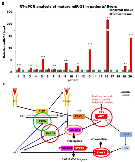
MiR-21 expression shows a direct correlation with activation of PI3K, TGF-β and STAT3 (PTS) signaling network proteins in LmiR21 zebrafish and human nonviral hepatocellular carcinomas (HCC). (A) Expression patterns of Ptenb, p-Akt, Smad7, p-Smad3 and p-Stat3 in 10 mpf LmiR21 ± Dox liver samples after immunohistochemical (IHC) staining. Scale bar: 50 μm. Positive signal areas per field were quantified. (B) Western blot images (left) and quantitative data (right) of PTS signaling regulatory protein in 10 mpf LmiR21 ± Dox liver. (C) Human nonviral HCC samples (n = 20) were subjected to IHC of PTEN, p-AKT, SMAD7, p-SMAD3 and p-STAT3 expression. Scale bar: 50 μm. (D) miR-21 expression profiles in the surrounding normal and liver cancer tissues from nonviral HCC patients (n = 20) were analyzed by RT-qPCR. Positive correlation of miR-21 with PTS signaling regulatory genes in 60% of the nonviral HCC samples were observed. (E) Proposed mechanism by which miR-21 links the NAHCC development. MiR-21 mediates a newly discovered PI3K/Akt, TGF-β/Smad3 and Stat3 (PTS) signaling loop and feed forward loop involving Smad3/Stat3 dependent on miR-21 expression. (Abbreviations: →: activated, ─●: inhibited) Statistically significant differences from LmiR21 − Dox were denoted by * (p < 0.05), ** (p < 0.01) and *** (p < 0.001) for panels A, B and D.

Expression Data

Expression Detail
Antibody Labeling
Phenotype Data

Phenotype Detail
Acknowledgments
This image is the copyrighted work of the attributed author or publisher, and ZFIN has permission only to display this image to its users. Additional permissions should be obtained from the applicable author or publisher of the image. Full text @ Cancers