
Fig. 2
- ID
- ZDB-FIG-210226-8
- Publication
- Westerich et al., 2020 - Bioorthogonal mRNA labeling at the poly(A) tail for imaging localization and dynamics in live zebrafish embryos
- Other Figures
- All Figure Page
- Back to All Figure Page
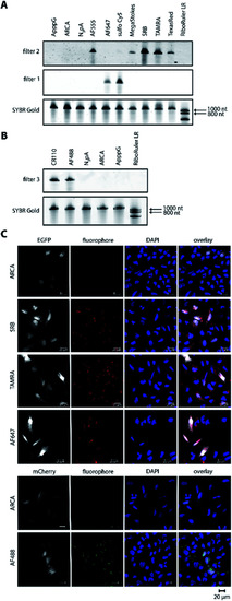
Bioorthogonal labeling of mRNAs at the poly(A) tail with different fluorophores enables their visualization in HeLa cells. (A) PAGE analysis of egfp mRNAs labeled with indicated fluorophores or controls (ApppG, ARCA, N3pA). (B) PAGE analysis of mcherry mRNAs labeled with indicated DBCO-fluorophores or controls. (7.5% PA gel, 20 W, 2.5 h, rt; filter 1: ≥575 nm, filter 2: ≥665 nm, filter 3: ≥510 nm). (C) Confocal microscopy of HeLa cells transfected with egfp or mcherry mRNA with indicated labels. Signals (red or green dots) from the fluorophores, EGFP/mCherry and DAPI channels enabled visualization of mRNA, EGFP/mCherry and nuclei, respectively. Scale bar = 20 μm. |