|
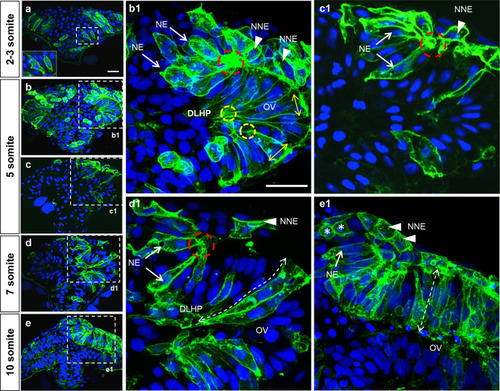
Cell shape changes in the deep layer of the ANP that contribute to DLHP and neural fold formation.a–e Transverse section, at the level of the forebrain, of embryos at the 2–3 (a), 5 (b, b1, c, c1), 7 (d, d1), and 10 (e, e1) somite stages mosaically expressing mGFP (green) and labeled with the nuclear marker DAPI (blue). The inset in a is a higher magnification of dashed area in a. b1–e1 Higher magnifications of regions delineated by dotted lines in b–e. Annotations: red dashed circle = basal constriction of NE component of neural fold; yellow circle = apical constriction of DLHP cell; arrows = NE component of neural fold; arrowheads = NNE component of neural folds; double dashed arrow = elongated deep cells of the optic vesicle, asterisks = dividing cells in the prospective telencephalon. Scale bars: 25 μm in a and b1.
|