
Figure 5
- ID
- ZDB-FIG-210128-99
- Publication
- Takaki et al., 2020 - Schistosoma mansoni Eggs Modulate the Timing of Granuloma Formation to Promote Transmission
- Other Figures
- All Figure Page
- Back to All Figure Page
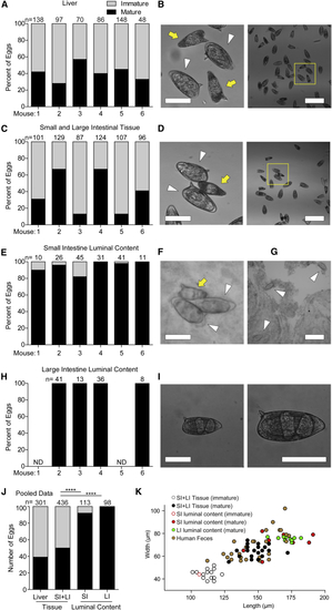
Mature Eggs Translocate into the Lumen of the Intestines (A–I) Quantification (A, C, E, and H) and representative brightfield images (B, D, F, G, and I) of mature and immature eggs found in the liver (A and B), small and large intestinal wall tissue and vasculature (C and D), small intestinal luminal content (E–G), and large intestinal luminal content (H and I) for six individual (B and D) Representative images with image (left) showing immature (yellow arrow) and mature (white arrowhead) magnified from yellow square in wide-field image (right). (F and G) Images of eggs from the lumen of the small intestine, showing two mature eggs in contact with one immature egg (F), and a wide-field image showing three mature eggs (G). (I) Representative image of an egg recovered from feces at low resolution (left) and higher resolution with developed miracidia visible (right). (J) Pooled data for mice 2, 3, 4, and 6 from (A, C, E, and H). SI, small intestine; LI, large intestine. (K) Dimensions of eggs from this experiment that were classified as immature or mature (open or closed circles, respectively) plotted with eggs shed in the feces of |