
Figure 4
- ID
- ZDB-FIG-201208-29
- Publication
- Baek et al., 2020 - An Embryonic Zebrafish Model to Screen Disruption of Gut-Vascular Barrier upon Exposure to Ambient Ultrafine Particles
- Other Figures
- All Figure Page
- Back to All Figure Page
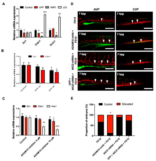
UFP exposure down-regulates mRNA expressions of endothelial tight junction (TJ) protein and the Notch target gene to disrupt the GVB. mRNA expressions of TJ proteins, including zonula occludens1 (Zo1), claudin 1 (Cldn1), and occludin 1 (Ocln1), and the Notch target genes, including Hairy and enhancer of split-1 (Hes1), were assessed in vitro by cultured human aortic endothelial cells (HAEC). (A) UFP exposure (25 μg·mL−1 for 6 h) inhibited Zo1 and Cldn1 mRNA expressions, whereas Ocln1 mRNA remained unchanged. While Iwr1 treatment (10 μM) diminished overall TJ mRNA expression, LiCl (20 mM) up-regulated Cldn1 and Ocln1 mRNA expression (* p < 0.05 vs. DMSO for Iwr1, H2O for LiCl, n = 3, ** p < 0.01, *** p < 0.001). (B) UFP exposure (25–50 μg·mL−1 for 6 h) down-regulated both TJ (Zo1 and Cldn1 mRNA) and Notch target genes (Hes1) mRNA expression in a dose-dependent manner ( |