FIGURE

Fig. 1

ID
ZDB-FIG-201113-22
Publication
Cook et al., 2020 - Visualisation of cholesterol and ganglioside GM1 in zebrafish models of Niemann-Pick type C disease and Smith-Lemli-Opitz syndrome using light sheet microscopy
Other Figures
All Figure Page
Back to All Figure Page
Fig. 1

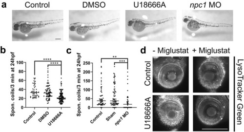
Identification of NPC phenotypes in a Npc1 inhibitor and npc1 morpholino zebrafish larvae model. NPC phenotypes were induced in the zebrafish larvae using either the Npc1 inhibitor, U18666A (2 µg/ml), or npc1 morpholino (npc1 MO) injected at the 1–2 cell stage with DMSO and Sham controls as necessary; a Morphology of 48 h post fertilisation (hpf) zebrafish by light microscopy. Quantitative behavioural analysis of 24 hpf zebrafish measuring number of spontaneous coils over 3 min in 2 µg/ml U18666A treated (b) and npc1 morphant (c) zebrafish, *p < 0.05, **p < 0.01, ****p < 0.0001, Kruskal–Wallis test with Dunn’s post-hoc correction. Representative images of LysoTracker Green staining of 96 hpf control and U18666A treated zebrafish larvae eyes with and without 500 µM miglustat co-treatment (d). Scale bars = 280 µm. N = 3–5, with a minimum of 10 fish per N

Expression Data

Expression Detail
Antibody Labeling
Phenotype Data
Fish:
Conditions:
Knockdown Reagent:
Observed In:
Stage Range: Prim-5 to Day 4

Phenotype Detail
Acknowledgments
This image is the copyrighted work of the attributed author or publisher, and ZFIN has permission only to display this image to its users. Additional permissions should be obtained from the applicable author or publisher of the image. Full text @ Histochem. Cell Biol.