|
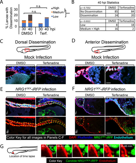
Blockade of blood flow partially limits dissemination.(A-G) Tg(fli1:EGFP) larvae, with GFP-expressing endothelial cells, were infected as previously described, treated with vehicle (DMSO) or terfenadine (2 μM) and processed for histology at 40 hpi. (A-B) Percent larvae with dissemination at 30 and 40 hpi scored as having “low” (1–10), “medium” (10–50), and “high” (>50 yeast) disseminated yeast. Pooled from 7 experiments, DMSO n = 110, Terfenadine n = 118. Fisher’s Exact test was used to test for differences between “low” and “medium” plus “high” scores at 40 hpi, *** p ≤ 0.001. (C-F) Sections of Tg(fli1:EGFP) mock infected (C-D) and Candida-infected (E-F) larvae compare dorsal dissemination (E) and anterior dissemination (F) events to the same tissue in mock-infected fish. Sections were stained with DAPI (nuclei, red) to indicate surrounding cells and phalloidin (actin; blue) to indicate host structures. NRG1OEX-iRFP (green) and Tg(fli1:EGFP) (cyan) retained fluorescence during sectioning and staining. White asterisks indicate disseminated yeast which appear to be embedded in the yolk syncytial layer and larval heart. Scale bar = 100 μm. (G) A vehicle-treated Tg(fli1:EGFP) (red) fish infected with NRG1OEX-iRFP (green) with intact blood flow was imaged by time-lapse. Area in red box is the focus of the time-lapse, with frames taken from S14 Movie a cropped area of the complete time lapse included as S15 Movie. Scalebar = 10 μm.
|