
Fig. 1
- ID
- ZDB-FIG-200818-15
- Publication
- Begovich et al., 2020 - Phosphoribosyl pyrophosphate synthetase polymerization influences lens fiber organization in zebrafish
- Other Figures
- All Figure Page
- Back to All Figure Page
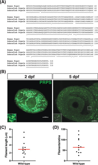
Phosphoribosyl pyrophosphate synthetase (PRPS) forms filaments in the retina at 5 days post fertilization (dpf). A, Protein sequence alignment (UniProt) of human PRPS (P60891) to zebrafish PRPS paralogs Prps1a (Q4KME9) and Prps1b (Q08CA5). Human PRPS1 has ~96% identity to both zebrafish paralogs. B, Antibody raised against human PRPS1 recognizes filaments at 5 dpf in the zebrafish eye. Representative maximum intensity projections of confocal images depict left eyes at 2 and 5 dpf stained for PRPS; rostral is to the left and dorsal is to the top. We note that we have never detected filament-like staining in the wild-type eye using the goat anti-rabbit secondary antibody in the absence of the anti-PRPS antiserum. Insets in bottom left corners show zoomed views of boxed areas. Scale bars: 50 μm. (n = 12 embryos for each stage). C, Graph plots the average length of the PRPS filaments observed in each eye examined at 5 dpf. Red line indicates the median value. D, Graph plots the number of PRPS filaments found in each eye examined at 5 dpf. Red line indicates the median value |
| Genes: | |
|---|---|
| Fish: | |
| Anatomical Term: | |
| Stage: | Day 5 |