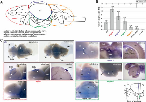
alpl expression pattern in whole adult zebrafish brains. (A) Scheme of different brain regions used for qPCR. (B) alpl expression at the mRNA levels determined by qPCR targeting exon 5/6 in different regions of adult zebrafish brain. Statistics were calculated using two-sided student t-test, paired; **p < 0.01, *p < 0.05, n.s., not significant; ref, reference. (C) ISH visualized expression domains of alpl in whole adult brains (marked by black and white arrows). Signals were detected in the telencephalon, tectum opticum and hypothalamus. Vibratome cross sections of single regions showed tissue-internal alpl expression. Control samples were hybridized with sense probe instead of antisense probe. Scale bar: 250 µm (whole brain images); 100 µm (close up view and sections). CCe, corpus cerebelli; Dl/Dd, lateral/dorsal zone of telencephalon; Hd, dorsal zone of the periventricular hypothalamus; IL, inferior lobe of hypothalamus; LR, lateral recess of diencephalic ventricle; Mo, medulla oblongata; PG, preglomerular area; PGZ, peripheral growth zone (layer 1 and 2); PiT, posterior inferotemporal cortex; Tel, telencephalon; TeO, tectum opticum; TH, tuberal hypothalamus; Val, ventral anterior thalamic nucleus; lateral part; Vam, ventral anterior thalamic nucleus; medial part.
|
