|
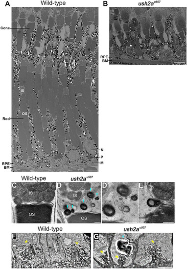
Retinal ultrastructure of ush2au507 zebrafish. Transmission electron microscopy was used to examine the wt and ush2au507 retinal ultrastructure at 6 months post fertilization (mpf). The wt retina showed tiers of morphologically distinct cone and rod photoreceptors with overlying retinal pigment epithelium (RPE) with long apical projections that interdigitated with the photoreceptors (A). Among mostly preserved tissue, regions of rod loss could be observed in the ush2au507 retina (B). Presumptive lysosomal structures (blue arrows) were noted at the inner and outer segment boundary (D, E) and at the synapses (G) of the ush2au507 photoreceptors at 6 mpf. Ribbon synapses are indicated by *. These vesicles were not observed in wt photoreceptors (C, F). IS, inner segment; OS, outer segment; IS, inner segment; OS, outer segment; RPE, retinal pigment epithelium; BM, Bruch’s membrane; N, nucleus; P, phagosome; M, melanosome. Scale bars = 5 μm (A, B, F, G) and 500 nm (C–E).
|