FIGURE

Figure 4—figure supplement 1.

ID
ZDB-FIG-200530-8
Publication
Williams et al., 2020 - Nodal and Planar Cell Polarity signaling cooperate to regulate zebrafish convergence and extension gastrulation movements
Other Figures
All Figure Page
Back to All Figure Page
Figure 4—figure supplement 1.

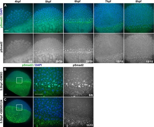
Nodal signaling activity in intact embryos.

(A) Representative confocal z-projections of immunofluorescent (IF) staining for phosphorylated Smad2 (bottom) overlaid with DAPI-labeled nuclei (top) in WT embryos fixed at the time points indicated. (B) Representative confocal z-projections of pSmad2 IF and DAPI staining in 5.5 hpf WT embryos treated with DMSO (top) or SB505124 (bottom), starting at 3 hpf. Panels to the right are magnified views of the regions within the white squares. Fractions indicate the number of embryos with the depicted phenotype over the total number of embryos examined for each condition and/or timepoint. The animal pole is at the top of all images. Scale bars are 50 μm.

Expression Data

Expression Detail
Antibody Labeling
Phenotype Data

Phenotype Detail
Acknowledgments
This image is the copyrighted work of the attributed author or publisher, and ZFIN has permission only to display this image to its users. Additional permissions should be obtained from the applicable author or publisher of the image. Full text @ Elife