
Fig. 6
- ID
- ZDB-FIG-200518-39
- Publication
- Lu et al., 2020 - Reissner fibre-induced urotensin signalling from cerebrospinal fluid-contacting neurons prevents scoliosis of the vertebrate spine
- Other Figures
- All Figure Page
- Back to All Figure Page
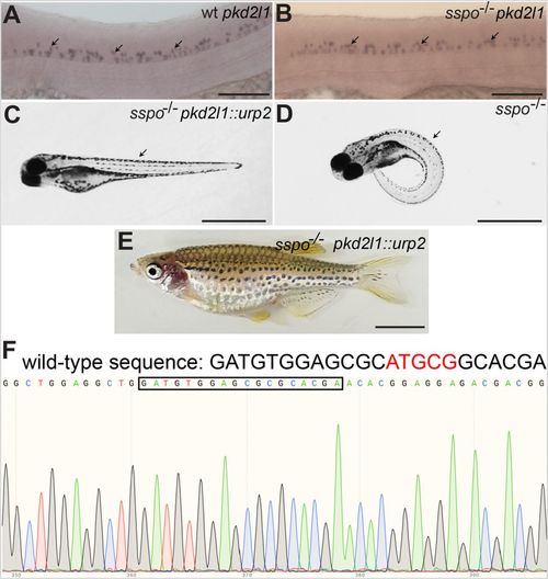
Axial defects of sspo mutants can be rescued by restoring urp2 expression specifically in CSF-cNs using the pkd2l1 promoter. (A) A wild-type embryo, showing pkd2l1 expression in CSF-cNs (arrows). (B) An sspo-mutant embryo showing normal levels and pattern of pkd2l1 expression in CSF-cNs (arrows). Embryos depicted in A and B were at 26 hpf and are representative of a minimum of 25 embryos analysed for each genotype. Scale bars: 100 μm. (C) An sspo-mutant larva rescued of axis curvature (arrow) by restoration of urp2 expression using the pkd2l1::urp2 transgene. A total of 150 embryos with straight axes from sspo/+ in-cross injected with pkd2l1::urp2 transgene from two independent experiments were genotyped, and 20 were found to be sspo-homozygous mutants. (D) An sspo-mutant larva. Note the strong ventrally curved axis (arrow). The larvae depicted were at 5 dpf. Scale bars: 1 mm. (E) An sspo-mutant adult rescued of its scoliotic spine by restoration of urp2 expression using the pkd2l1::urp2 transgene. Of 92 adults with straight axes, 31 were wild type, 60 were heterozygous and one was a homozygous mutant. Scale bar: 0.5 cm. (F) Electropherogram showing homozygous mutant genotype of the rescued sspo-mutant adult depicted in E. The wild-type sequence is indicated on top, with the five bp that are deleted in the mutant in red. The mutant sequence (with five bp deletion) is highlighted with the black box. |