FIGURE

Figure 5

ID
ZDB-FIG-200306-30
Publication
Pang et al., 2020 - Mutant dlx3b disturbs normal tooth mineralization and bone formation in zebrafish
Other Figures
All Figure Page
Back to All Figure Page
Figure 5

Surfaces of the second and fourth ventral pharyngeal teeth of the <italic>dlx3b</italic> homozygous mutation zebrafish exhibited abnormal pits.

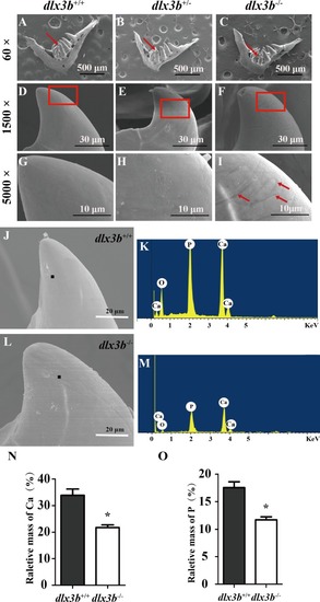
(A–I) SEM images showing the surface of the second pharyngeal tooth. The red arrows showed the teeth we studies below in a 60× image (A–C). The fourth ventral pharyngeal teeth (red arrows in A–C) was magnified as images D–F. The red rectangle areas in D–F were then magnified as G–I. The red arrows (I) indicate the pits on the surface of the second ventral pharyngeal tooth of the dlx3b−∕− fish. n=10 for each group (J–M) Lower relative mass fractions of mineral elements in the dlx3b−∕− mutant. The scanning areas for energy spectrum analysis were selected on the cusp of the pharyngeal teeth showed as images J and L. Energy spectrum of zebrafish pharyngeal teeth was showed as images K and M, mainly including O, Ca, and P. (N–O) The relative mass fraction of Ca (N) and P (O) significantly decreased in the dlx3b−∕− zebrafish compared with those in their wild-type siblings. n = 5, P < 0.001.

Expression Data

Expression Detail
Antibody Labeling
Phenotype Data
Fish:
Observed In:
Stage: Adult

Phenotype Detail
Acknowledgments
This image is the copyrighted work of the attributed author or publisher, and ZFIN has permission only to display this image to its users. Additional permissions should be obtained from the applicable author or publisher of the image. Full text @ Peer J.