FIGURE

Figure 3

ID
ZDB-FIG-200306-28
Publication
Pang et al., 2020 - Mutant dlx3b disturbs normal tooth mineralization and bone formation in zebrafish
Other Figures
All Figure Page
Back to All Figure Page
Figure 3

Body curvature of <italic>dlx3b</italic> homozygous mutant zebrafish.

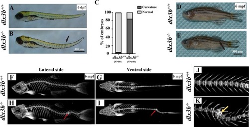
(A–B) Body curvature of 6–7 dpf larvae under a stereomicroscope. The body curvature in dlx3b−∕− (B) initially occurred in the early juvenile stages and was mainly limited to the distal end of the tail (black arrow), while the wild type (A) was showed no curvature. Scale bar: 300 µm. (C) Relative frequencies of dorsal curvature phenotypes in dlx3b+∕+ and dlx3b−∕− embryos. n = 100 for each group. (D–E) Mutant adult zebrafish (D) exhibited a severe curvature of the spine compared with wild-type siblings (E), especially in the tail part (black arrow). (F–I) Micro-CT results showing the spine from the lateral side (F & H) and ventral side (G & I). The abdominal and caudal spine of the dlx3b−∕− zebrafish developed a marked curvature in both the dorsal–ventral (G&I) and medial–lateral planes (F&H, red arrows). (J–K) Magnified imaging showing vertebral hypoplasia in the dlx3b−∕− zebrafish. A radiopaque area was observed in the caudal spine region (K, yellow arrow).

Expression Data

Expression Detail
Antibody Labeling
Phenotype Data
Fish:
Observed In:
Stage Range: Day 6 to Adult

Phenotype Detail
Acknowledgments
This image is the copyrighted work of the attributed author or publisher, and ZFIN has permission only to display this image to its users. Additional permissions should be obtained from the applicable author or publisher of the image. Full text @ Peer J.