
Fig. S3
- ID
- ZDB-FIG-190827-22
- Publication
- Santos et al., 2019 - Exogenous WNT5A and WNT11 proteins rescue CITED2 dysfunction in mouse embryonic stem cells and zebrafish morphants
- Other Figures
- All Figure Page
- Back to All Figure Page
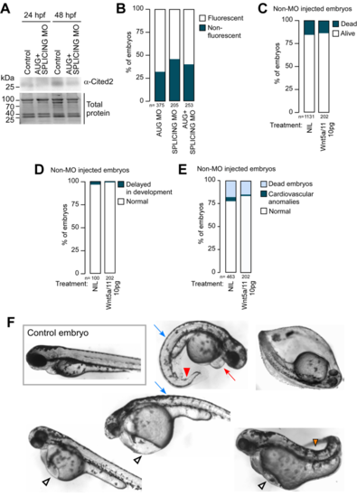
Cited2 morphants display developmental defects, but the sole microinjection of recombinant Wnt5a and Wnt11 does not affect the development of normal embryos. (A) Top panel: Cited2 protein levels determined by western blotting in protein extracts from zebrafish embryos at 24 hpf and 48hpf after microinjection of either combined AUG and SPLICING (AUG+SPLICING MO, 2.5 ng of each morpholino) or control non-injected embryos (Control). Bottom panel: Total protein stained with TGX Stain-Free™ FastCast™ Acrylamide Kit (Bio-Rad) transferred on the PVDF membrane was used to control for loading. The position of the Cited2 protein is in agreement with the predicted molecular weight (~25 kDa). (B) Percentage of fluorescent embryos at 6 hpf and after the individual and combined microinjection of AUG MO (5 ng) and SPLICING MO (5ng) (AUG+SPLICING MO, 2.5 ng of each morpholino) at 1-cell stage. (C) Percentage of dead and live embryos at 24 hpf and after combined microinjection of rWnt5a and rWnt11 (5 pg of each protein) or no injection at 1-cell stage. (D) Percentage of live embryos presenting developmental delays at 24 hpf and after microinjection of rWnt5a and rWnt11 as described in B. (E) Percentage of dead and live embryos presenting a normal morphology or cardiac anomalies at 72 hpf and after microinjection of rWnt5a and rWnt11 as described in B. For all panels, n represents the number of embryos analysed in each condition in at least 2 independent experiments. (F) Brightfield images of live embryos showing the representative morphology at 72 hpf of control embryos and embryos injected with 5 ng of anti-Cited2 morpholinos as described in Figure 4B. The red arrow indicates pericardial edema, the blue arrow an abnormal curvature of the spine, the white arrowhead a slight swelling or edema of the of the yolk sac, the red arrowhead a curvy tail and the orange arrowhead a severe notochord defect. An embryo that fail to hatch is also represented. |
| Fish: | |
|---|---|
| Knockdown Reagents: | |
| Observed In: | |
| Stage: | Protruding-mouth |