
Fig. S3
- ID
- ZDB-FIG-190819-11
- Publication
- Veloso et al., 2019 - Dephosphorylation of HDAC4 by PP2A-Bδ unravels a new role for the HDAC4/MEF2 axis in myoblast fusion
- Other Figures
- All Figure Page
- Back to All Figure Page
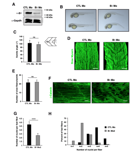
Knockdown of PP2A-Bδ in zebrafish embryos. (A) Western blot analysis of Bδ (α-Bδ) in 48 hpf zebrafish embryos injected with control morpholino (CTL Mo) and a morpholino targeting zebrafish Bδ orthologue (Bδ Mo). GAPDH (α-GAPDH) was used as loading control. Images are representative experiments from 2 independent experiments. (B-E) (B) Bright field pictures representative of at least 10 independent experiments, (C) quantification of somite angles (γ; nc: notochord), (D) visualization of slow twitch myotome by confocal microscopy and staining for slow skeletal myosin (green) and (E) quantification of slow fibers in each somite of 48 hpf zebrafish embryos injected with control morpholino (CTL Mo, n=8 for (C,D) and n=10 for (E)) or a morpholino against Bδ (Bδ Mo, n=17 for (C,D) and n=9 for (E)). Unpaired two-tailed t-test, *** P<0.001, ns: not significant. (F-H) (F) Confocal pictures of β-catenin (green) highlighting striations, (G) quantification of the number of nuclei per fiber and (H) proportion of fibers with the indicated number of nuclei in fast myofibers of control (CTL Mo, n=8 for (F) and n=5 for (G,H)) or PP2A-Bδ (Bδ Mo, n=17 for (F) and n=7 for (G,H)) morphant embryos. Unpaired two-tailed t-test, **** P<0.0001, ns: not significant. |