
Fig. S4
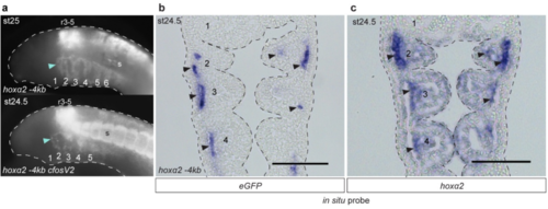
GFP reporter expression driven by lamprey hoxα2 upstream regions in transient transgenic lamprey embryos. a, Lateral views of st24-25 transient transgenic lamprey embryos showing GFP expression in rhombomeres (r), somites (s) and NC of the pharyngeal arches (numbered), driven by the hoxα2 -4kb enhancer with or without the mouse c-Fos minimal promoter. In cloning the c-Fos promoter between the lamprey enhancer and the GFP coding sequence, two alternative reporter constructs were generated: hoxα2 -4kb cFosV1 with the upstream lamprey sequence fully intact, hoxα2 -4kb cFosV2 with the 5’UTR partially removed. In both cases, the insertion of the c-Fos promoter increased levels of reporter expression but did not influence tissue-specific expression domains. GFP-expressing embryos shown are representative of the expression potential of the reporter construct in each case, as inferred from screening many (typically more than 100) injected embryos (see Supplementary Table 2 for expression statistics). b, Frontal section through a transient transgenic lamprey embryo at st24.5, revealing GFP transcripts in the NC-derived mesenchyme (arrows) of the pharyngeal arches (numbered), driven by hoxα2 -4kb. c, Frontal section through a st24.5 lamprey embryo showing endogenous hoxα2 expression. Arrows indicate elevated expression in the NC-derived mesenchyme. The pharyngeal arches are numbered. Scale bars: 100μm. Abbreviations: r, rhombomere; s, somites. |