
Fig. 3
- ID
- ZDB-FIG-190730-24
- Publication
- Kolvenbach et al., 2019 - Rare Variants in BNC2 Are Implicated in Autosomal-Dominant Congenital Lower Urinary-Tract Obstruction
- Other Figures
- All Figure Page
- Back to All Figure Page
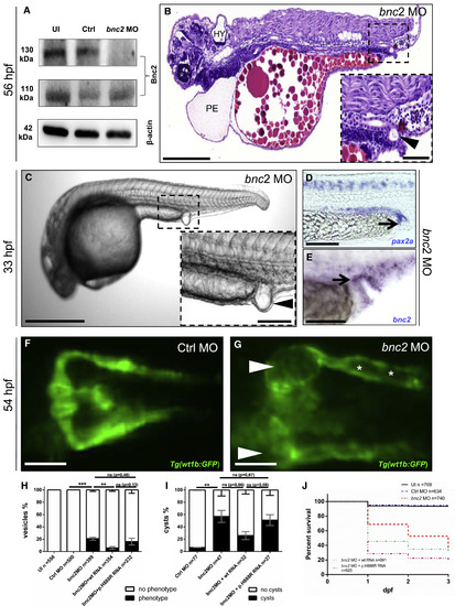
Depletion of Bnc2 Causes Pericardial Effusion, Hydrocephalus, Glomerular Cysts, and Distal Pronephric-Outlet Obstruction in Zebrafish (A) Immunoblot analysis shows a protein decrease in bnc2 MO-injected zfl for Bnc2-202 (130 kDa, C7DZJ6 according to UniProt ID) but not for Bnc2-201 (110 kDa, F1R42 according to UniProt ID) at 2 days dpf, which is as predicted for the specific MO target side. UI = uninjected. (B) H&E staining of a sagittal section of a MO-injected zfl (lateral view, head to the left) shows (PE), hydrocephalus (HY), abnormal body curvature and a “vesicle” due to an outlet obstruction of the pronephric ducts (arrowhead in enlargement in B) at 56 hpf. (C) Zfl injected with bnc2 MO frequently develop a distal-outlet obstruction at 33 hpf; this obstruction is highlighted by an arrowhead in the enlargement of (C). (D) Whole-mount ISH with a pax2a probe relates the constituent parts of the bnc2 MO-induced pronephric-outlet obstruction to distal parts of the pronephric ducts and the cloacal region (black arrow). (E) ISH with a bnc2 probe in bnc2 MO-injected embryos shows bnc2 mRNA expression in relevant tissues forming a pronephric-outlet obstruction (black arrow). (F and G) Zfl injected with bnc2 MO develop glomerular cysts (white arrowheads in G) and dilatation of the pronephric ducts (white asterisk in G). Images from in vivo observation through fluorescence microscopy (dorsal view) in Tg(wt1b:GFP) were taken at 54 hpf in zfl injected with control (Ctrl) MO (F) and bnc2 MO (G). Tg = transgenic zebrafish line. (H) The graph shows 100% of all at 1 dpf surviving zfl of the five different cohorts: UI, Ctrl MO, bnc2 MO, bnc2 MO + WT human RNA, and bnc2 MO + mutated human RNA (transcript ENST00000380672) bearing the p.His888Arg variant (for absolute numbers, see Figure 3H). A distal “vesicle” due to an outlet obstruction (phenotype) of the pronephric ducts can be seen in 21% of bnc2MO-injected zfl compared to 0% of zfl with a “vesicle” in both control groups (p < 0.05, unpaired t test). 6% of zfl injected with bnc2 MO + WT mRNA develop a “vesicle,” and so do 16% of zfl injected with bnc2 MO + p.H888R. Data are presented as means with standard error of the mean (SEM). (I) Quantification of glomerular cyst rates (phenotype) in Tg(wt1b:GFP) zfl at 2 dpf depicts significantly (p < 0.05, unpaired t test) clear glomerular cysts in 57% of bnc2 MO-injected zfl. 26% of zfl injected with bnc2 MO + WT RNA develop pronephric cysts; 51% of those injected with bnc2 MO + p.His888Arg RNA develop pronephric cysts. Data are presented as means with SEM. (J) Quantification of death rates at up to 3 dpf shows an increased mortality up to 64% in bnc2 MO-injected zfl compared to zfl injected with UI (7%) and Ctrl MO (6%). With bnc2 MO injected, zfl show significantly (p < 0.0001, Mantel-Cox test) reduced survival. Co-injection of bnc2 MO + human WT RNA results in a mortality of 69%. Aggravated mortality up to 81% can be detected in the zfl injected with bnc2 MO + p.His888Arg RNA. Scale bars represent 200 μm (B), 50 μm (magnification in B), 500 μm (C), 100 μm (magnification in C), and 100 μm (D–G). ∗∗p < 0.005, ∗∗∗ p < 0.0005. |
| Gene: | |
|---|---|
| Fish: | |
| Knockdown Reagent: | |
| Anatomical Terms: | |
| Stage: | Prim-15 |
| Fish: | |
|---|---|
| Knockdown Reagent: | |
| Observed In: | |
| Stage Range: | Prim-15 to Protruding-mouth |