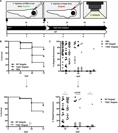
Emergency granulopoiesis mediates long-term host defense. (A) Schematic of reinfection assays. At 2 dpf, wild-type (WT) AB zebrafish larvae were injected in the HBV with PBS or a low dose (0.5 × 10³ to 2.0 × 10³ CFU) of GFP+S. flexneri M90T. At 4 dpf, i.e., 48 h post-primary injection (hp1i), all larvae were injected with a high dose (>2.0 × 10⁴ CFU) of mCherry-expressing (mCherry+) S. flexneri M90T. Analyses were performed on larvae up to 72 h post-secondary infection (hp2i). (B and C) WT AB larvae were injected with PBS (open circles) or “primed” with wild-type or T3SS− GFP+S. flexneri M90T (closed circles), prior to a high dose of mCherry+S. flexneri M90T at 48 hpi, as described above. (B) Survival curves pooled from 4 independent experiments using n ≥ 9 larvae per condition per experiment. Up to three larvae per condition were taken for CFU at the 24 and 48 h time points. The top graph represents collated data. The bottom graph represents only Shigella-primed larvae, a subset of the above data. The P value between conditions was determined by log-rank Mantel-Cox test. Significance was defined as P < 0.05 (*). (C) Fluorescent mCherry+S. flexneri M90T burden of larvae was imaged by stereomicroscopy over time, and images were analyzed to produce fluorescence intensity measurements (as in Fig. S2A). Data were pooled from 4 independent experiments with n ≥ 4 per time point per condition per experiment. The top graph represents collated data. The bottom graph represents only Shigella-primed larvae, a subset of the above data. P values between conditions at cognate time points were determined by unpaired two-tailed Student’s t test. Significance was defined as P < 0.05 (*).
|
