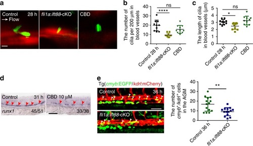
Blocking formation or function of primary cilia impairs hematopoietic stem and progenitor cell (HSPC) development. a Confocal imaging of cilia in the aorta-gonad-mesonephros (AGM) region in kdrl:mCherry/βact:Arl13b–GFP double-transgenic line with fli1a:ift88-cKO-functional injection, or ciliobrevin D (CBD) treatment at 28 hpf. White arrow denotes the blood flow direction. Scale bar, 5 µm. b, c The quantification of primary cilia number and length in the blood vessels of AGM in a. Data in b, c represent the analysis results of one-way ANOVA–Dunnett test. Error bars, mean ± s.d., n = 10 embryos. ns non-significant, *P < 0.05, ****P < 0.0001. d WISH result of runx1 (red arrowheads) in control and CBD-treated embryos at 31 hpf. The red arrowheads indicate the expression of HSPC marker runx1. Scale bar, 100 µm. e The kdrl:mCherry+/cmyb:EGFP+ HE cells (white arrowheads) in the AGM region in fli1a:ift88-cKO-injected embryos (left panel) with quantification (right panel) at 36 hpf. Cmlc2:EGFP-negative embryos were used as a negative control (control). Scale bar, 50 µm. Error bars, mean ± s.d., n = 14 embryos. **P < 0.01, Student’s t-test
|
