
Fig. 6
- ID
- ZDB-FIG-190627-30
- Publication
- Campo-Paysaa et al., 2019 - Generation of the squamous epithelial roof of the 4th ventricle
- Other Figures
- All Figure Page
- Back to All Figure Page
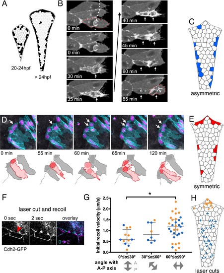
(A) Schematics of roof plate that cumulatively map the distribution of cell divisions between 20 and 24 hpf and from 24 to 36 hpf from 125 embryos. (B) Time-lapse sequence of veil cell (outlined by red dots) during an asymmetric division (see also Figure 6—video 1) with cleavage plane parallel to roof plate border (dotted white line). Two timepoints in mitosis indicated by single white arrow at 30 and 35 min. Position of two daughters indicated by white arrows from 40 min onwards. Medial daughter becomes a squamous roof plate cell (marked S) and lateral daughter becomes a veil cell (marked V and outlined by red dots). Images also contain a second veil cell and an established squamous cell that have been indicated with reduced brightness in the drawing. (C) Schematic map of location of 26 asymmetrically fated veil cell divisions. (D) Five frames from time-lapse of a symmetrically fated veil cell division starting at 23 hpf that generates daughters with same fates (two veil cells). Arrows indicate mother and daughter veil cells. One daughter inherits the original basolateral veil while the other rapidly regenerates a veil after mitosis (see also Figure 6—video 2). (E) Schematic map of 19 symmetrically fated veil cell divisions. (F) Time-lapse sequence (see also Figure 6—video 3) showing laser cut of a cell-cell junction in the roof plate of a Cdh2-GFP embryo at 24hpf. Site of laser ablation is indicted with a red arrowhead. Arrows indicate positions and recoil of adjacent tricellular vertices. (G) Quantification of initial recoil velocity after laser cut as a function of the angle to the anteroposterior axis shows a significantly higher velocity following parallel versus perpendicular cuts (Two-tailed Mann-Whitney, p=0.0113). Recoil in the region proximal to the upper rhombic lip (orange) is similar to that adjacent to the lower rhombic lip (blue). (H) Map of positions and angles of laser cuts. |