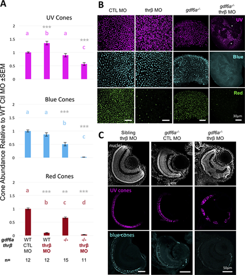
Knockdown of thrβ disrupts retinal lamination, including the photoreceptor layer, in gdf6as327/s327 mutants. (A) Thrβ knockdown with splice-blocking morpholino in gdf6as327/s327mutants (labeled gdf6a−/− or “−/−”) fails to increase UV cone abundance, instead causing near-total loss of blue and red cones. Values with matching letters are not significantly different (Kruskal-Wallis test with Mann-Whitney pairwise comparisons, gray asterisksindicate *P < 0.05, **P < 0.01, ***P < 0.001 relative to WT + control [CTL] MO controls). UV cone abundance decreased significantly, in contrast to thrβ knockdown in wildtype fish. (B) Gaps or “holes” can be seen in the photoreceptor layer of gdf6as327/s327; thrβ MO-treated whole-mount retinas (two holes indicated by asterisks in UV image). These holes are not seen in CTL MO-injected gdf6as327s327 mutants, nor in wild-type or heterozygous morphants. (C) Radial sections of 4 dpf gdf6as327/s327; thrβ MO-treated retinas show disrupted retinal lamination, with gaps in the photoreceptor layer that are occupied by cells of the inner nuclear layer, and cells disrupting the inner plexiform layer between the inner nuclear layer and ganglion cell layer (n = 9 embryos examined per group).
|
