
Fig. 6
- ID
- ZDB-FIG-190528-12
- Publication
- Blokhina et al., 2019 - The telomere bouquet is a hub where meiotic double-strand breaks, synapsis, and stable homolog juxtaposition are coordinated in the zebrafish, Danio rerio
- Other Figures
- All Figure Page
- Back to All Figure Page
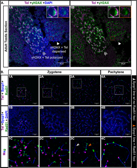
Double strand break (DSB) localization during male zebrafish meiosis I. A: Testis section stained for γH2AX (green), telomeres (Tel; magenta), and DNA (DAPI, blue). γH2AX staining is polarized when the telomeres are in bouquet formation (triple arrow, white boxes) and is more scattered when the telomeres are dispersed (arrow, orange boxes). Scale bar = 20 μm. B: Panels 1A-4C: Nuclear surface spreads stained for Sycp1 (blue), Rad51 (green), telomeres (Tel, magenta) and DNA (DAPI, blue); 1C-4C: Magnified (Mag) images are from regions indicated by white boxes in the top row panels. White arrowhead points to an example of an Sycp1 stretch with no Rad51 foci. Orange arrowhead points to an example of an Sycp1 stretch with Rad51 foci. 1A-4B scale bar = 5μm.
|
| Antibodies: | |
|---|---|
| Fish: | |
| Anatomical Terms: | |
| Stage: | Adult |