
Fig. 1
- ID
- ZDB-FIG-180913-38
- Publication
- Roovers et al., 2018 - Tdrd6a Regulates the Aggregation of Buc into Functional Subcellular Compartments that Drive Germ Cell Specification
- Other Figures
- All Figure Page
- Back to All Figure Page
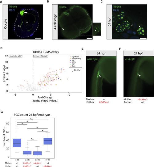
Tdrd6a Is Germline Specific and Required for PGC Formation (A) IHC for Tdrd6a in oocytes. Arrowhead and arrow indicate Tdrd6a staining in the nuage and Bb, respectively. Gray dashed line outlines the cell, n = nucleus. Scale bar, 10 μm. (B) IHC for Tdrd6a in 4-cell stage embryos. Arrows indicate Tdrd6a localization to the Gp. Scale bar, 100 μm. (C) Tdrd6a localizes to peri-nuclear nuage granules (arrowheads) in PGCs at 24 hpf. Scale bar, 7.5 μm. (D) MS of Tdrd6a IPs on an ovary, compared to IgG (immunoglobulin G) control. (E and F) 24 hpf embryos derived from wt (E) or tdrd6a mutant mothers (F) in a vasa:egfp background. Arrowheads indicate the PGCs. (G) Quantification of PGC numbers in 24 hpf embryos from the crosses indicated on the x axis (∗ indicates p value < 0.0001, n.s. = non-significant, calculated by Wilcoxon test). See also Figures S1 and S2. |
| Gene: | |
|---|---|
| Antibody: | |
| Fish: | |
| Anatomical Terms: | |
| Stage Range: | 4-cell to Adult |
| Fish: | |
|---|---|
| Observed In: | |
| Stage: | Prim-5 |
Reprinted from Developmental Cell, 46, Roovers, E.F., Kaaij, L.J.T., Redl, S., Bronkhorst, A.W., Wiebrands, K., de Jesus Domingues, A.M., Huang, H.Y., Han, C.T., Riemer, S., Dosch, R., Salvenmoser, W., Grün, D., Butter, F., van Oudenaarden, A., Ketting, R.F., Tdrd6a Regulates the Aggregation of Buc into Functional Subcellular Compartments that Drive Germ Cell Specification, 285-301.e9, Copyright (2018) with permission from Elsevier. Full text @ Dev. Cell