
Fig. 2
- ID
- ZDB-FIG-180829-11
- Publication
- Liu et al., 2018 - CRMP2 and CRMP4 Are Differentially Required for Axon Guidance and Growth in Zebrafish Retinal Neurons
- Other Figures
- All Figure Page
- Back to All Figure Page
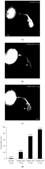
Knocking down CRMP2 induces growth defects of retinal axons. Morpholino was injected into the zygotes at the 1-2 cell stage. Embryos were allowed to grow until 4 days (4 days postfertilization, 4 dpf) and fixed with PFA. Lipophilic fluorescent dye DiI or DiD was injected into an eye of the larvae to label retinal axons. (a) An example showing that, in control MO-treated zebrafish larvae, retinal axons exit the eye, cross the midline, and grow into and arborize the whole tectum at 4 dpf. (b, c) Representative images of retinal axons of CRMP2 MO-treated embryos. Much less retinal axons grow into and arborized the tectum (white arrowheads) compared with that in control MO-treated embryos. (d) The growth defects of retinal axons induced by CRMP2 MOs are dose-dependent. The y-axis represents the percentage of eyes with growth defects of retinal axons. The doses of MOs are labeled under each column. The numbers in parentheses above each column indicate the amount of eyes. Scale bar: 50 μm. |
| Fish: | |
|---|---|
| Knockdown Reagent: | |
| Observed In: | |
| Stage: | Day 4 |