
Fig. 6
- ID
- ZDB-FIG-180827-37
- Publication
- Astone et al., 2018 - Zebrafish mutants and TEAD reporters reveal essential functions for Yap and Taz in posterior cardinal vein development
- Other Figures
- All Figure Page
- Back to All Figure Page
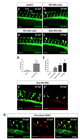
Yap1/Taz activity upregulation promotes vessel sprouting. (A–C) Confocal Z-stack projections of the midtrunk region of Tg(kdrl:GFP) 32 hpf embryos. (A) Representative image of a control injected embryo. (B,B’) Two TAZ-4SA mRNA injected embryos, showing an aberrant ISV sprout (arrowhead in B) and an anastomosis between adjacent ISVs (arrowhead in B’). (C) A mosaic embryo injected with pDestTol2CG2-fli1a-TAZ-4SA plasmid, showing three anastomosis between adjacent ISVs (arrowheads in C). (D,E) Quantification of the aberrant sprouting caused by Yap1/Taz activity upregulation. The number of non-anastomosed aberrant sprouts (D) and the number of anastomosis (E) between adjacent ISVs were evaluated. Both phenomena, observed in TAZ-4SA mRNA and pDestTol2CG2-fli1a-TAZ-4SA mosaic injected embryos, are extremely rare or absent at all in the controls. Controls: n = 49; TAZ-4SA mRNA-injected: n = 42; TEAD-VP16 mRNA-injected: n = 31; pDestTol2CG2-fli1a-TAZ-4SA plasmid-injected: n = 16. (F,F’) Tg(Hsa.CTGF:nlsmCherry)ia49/Tg(kdrl:GFP) double transgenic embryos injected with the pDestTol2CG2-fli1a:TAZ-4SA vector. A strong overactivation of the Hsa.CTGF reporter signal was observed in the nuclei of the endothelial cells undergoing anomalous sprouting (arrowheads in F) with respect to the other normal ISVs. (G) In mosaic embryos injected with the PCS2-fli1a:CAYAP-mKate plasmid a specific expression of the mKate was reported in conjunction with the anomalous endothelial sprouts (arrowhead). The plasmid is endothelium-specific, as highlighted by the co-localization (arrows) between the mosaic mKate and the GFP of the stable transgenic line Tg(kdrl:EGFP). Lateral view, anterior to the left, dorsal to the top. ***p < 0.001. ISV: intersegmental vessel; DLAV: dorsal longitudinal anastomotic vessel. Scale bar: 50 µm. |