
Fig. 2
- ID
- ZDB-FIG-180809-14
- Publication
- Svara et al., 2018 - Volume EM Reconstruction of Spinal Cord Reveals Wiring Specificity in Speed-Related Motor Circuits
- Other Figures
- All Figure Page
- Back to All Figure Page
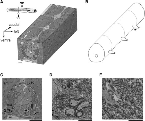
SBEM Volume of Zebrafish Spinal Cord (A) Three-dimensional EM volume comprising two complete segments from central spinal cord and two partial, adjacent segments. Schematic larval zebrafish shows the location of tissue sampling. (B) Schematic of spinal cord volume contained in the dataset. Dashed lines indicate approximate extent of spinal cord segments. Asterisk indicates the level of the pair of ventral roots from which MNs were reconstructed. (C) Transversal overview. am, axial musculature; ma, Mauthner axon; nc, notochord; sc, spinal cord; vr, ventral root. (D) Four large myelinated axons and many smaller unmyelinated axons exiting through a ventral root. Area as indicated in (C). (E) Densely packed neurites in the lateral neuropil. Area as indicated in (C). Scale bars: (C) 20 μm, (D) 2 μm, and (E) 1 μm. |