
Fig. 2
- ID
- ZDB-FIG-180720-2
- Publication
- Ji et al., 2018 - Directional selectivity of afferent neurons in zebrafish neuromasts is regulated by Emx2 in presynaptic hair cells
- Other Figures
- All Figure Page
- Back to All Figure Page
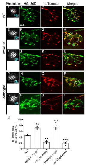
The branching pattern of a single afferent neuron in emx2 mutant neuromasts is altered. (A–D) A wildtype neuromast showing the seven A > P and eight P > A (red asterisks) HCs. All neurons are labeled green (HGn39D), while a single neuron is labeled red (neuroD:tdTomato). The fibers of the single tdTomato-positive neuron (C) overlap with a subset of the GFP-positive neurons (B) in the posterior part of neuromast (D). (E–L) Two emx2 ko neuromasts that contain only P > A HCs are shown (E,I). (E–H) The overlap between the single tdTomato-positive (G) and the GFP-positive neurons (F) in the neuromast is much broader than in wildtype (D). By contrast, the representation of tdTomato signals within the GFP-positive fibers of another emx2 ko neuromast (I–L) is less than the one in wildtype (D). (M–T) Both emx2 gof neuromasts (M, Q) show only A > P HCs, but one afferent neuron shows a broader distribution (M–P), whereas the other shows more restricted distribution of tdTomato in the neuromast (Q–T), when compared to the wildtype (D). Circles in (H,P) show fibers that are only GFP-positive. (U) Percentages of tdTomato-positive (single neuron) area per total GFP-positive (all neurons) area. The number of neuromasts: WT n = 3; emx2 ko major n = 3, minor n = 3; emx2 gof major n = 6, minor n = 4, the one-way ANOVA was used for the comparisons to WT. **p<0.01, ***p<0.001. Scale bar = 10 μm. |
| Fish: | |
|---|---|
| Observed In: | |
| Stage Range: | Long-pec to Day 6 |