
Fig. 4
- ID
- ZDB-FIG-180705-77
- Publication
- O'Connor et al., 2018 - MYO9A deficiency in motor neurons is associated with reduced neuromuscular agrin secretion
- Other Figures
- All Figure Page
- Back to All Figure Page
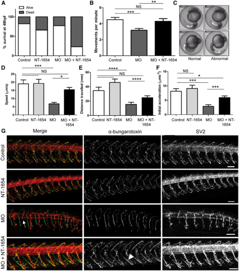
Effect of NT-1654 treatment in MYO9A-depleted zebrafish. (A) Graph depicting the survival rate of control, NT-1654 injected, MO injected and MO + NT-1654 injected zebrafish at 48 hpf (n = 100). (B) Movements performed by zebrafish within the chorion at 24 hpf in 1 min. Unpaired t-test, NS = not significant, **P ≤ 0.01, n > 40. (C) Example of chorion movement showing a full rotation of the zebrafish tail indicated by white arrows. (D) Speed, (E) distance and (F) initial acceleration of zebrafish swimming away following tactile stimulation at 48 hpf as calculated using TrackMate (ImageJ), n > 34. Unpaired t-test, NS = not significant, *P ≤ 0.05, **P ≤ 0.01, ***P ≤ 0.001, ****P ≤ 0.0001, error bars represent mean + standard error of the mean. (G) Immunofluorescent staining of 48 hpf zebrafish (control, NT-1654/MO/MO + NT-1654 injected) to visualize the neuromuscular junctions. SV2 stains pre-synaptic motor neurons and α-bungarotoxin detects the post-synaptic AChRs. MO injected fish display shortened axons (white arrow) and this is rescued by the application of NT-1654. AChR clusters also appear more prominent following addition of NT-1654 (white arrow head). Scale bars = 50µm. MO = morpholino, hpf = hours post-fertilization, AChRs = acetylcholine receptors, SV2 = synaptic vesicle protein 2. |
| Fish: | |
|---|---|
| Knockdown Reagents: | |
| Observed In: | |
| Stage Range: | Prim-5 to Long-pec |