
Fig. 5
- ID
- ZDB-FIG-180621-41
- Publication
- Green et al., 2017 - Recovery of shape and size in a developing organ pair
- Other Figures
- All Figure Page
- Back to All Figure Page
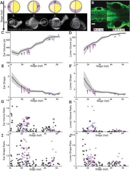
Localized UV treatment creates temporary asymmetry in ear and lumen size and shape. A: Oriented UV (302 nm) treatments. 6 hpf: yellow circles represent eggs (yolk, deep yellow; embryo, light yellow); UV orientation from Anterior, Posterior, Left or Right; purple crescents represent expected UV penetration. 24 hpf: embryos (oriented as 6 hpf; ears outlined white, lumens black) show damage near UV source (purple circles; note anterior UV often causes additional defects and early death). B: Early (26 hpf) and late (55.5 hpf) ear pairs for an individual (actb2:membrane‐citrine) UV‐treated on its left (same color used for same individual, C–J). Scale bar = 50 µm. Supplementary Movie S3 shows 3D visualizations of ear and lumen shapes and volumes. C–F: Ear (C) and lumen (D) volumes; ear (E) and lumen (F) shape ratios. Untreated wild‐type mean values (dark‐gray) and SD (light‐gray) as in Figure 3. Each arrow connects left (treated) and right ear values for one fish at one timepoint (three to four timepoints for three individuals, one color per individual). Squares replace arrows shorter than an arrowhead. G–J: B/S ratios for ear (G) and lumen (H) volumes; ear (I) and lumen (J) shape ratios (untreated wild‐type as in Fig. 3, gray dots). Black‐outlined colored dots match individuals in B–F. See Supplementary Table S2. |