
Fig. 3
- ID
- ZDB-FIG-180620-73
- Publication
- Murphy et al., 2018 - Placeholder Nucleosomes Underlie Germline-to-Embryo DNA Methylation Reprogramming
- Other Figures
- All Figure Page
- Back to All Figure Page
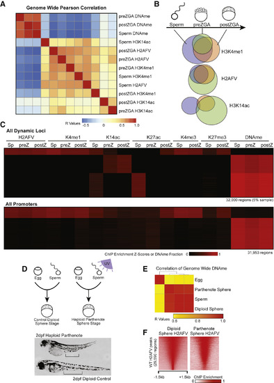
Maintenance of H2AFV and H3K4me1 at DNA Hypomethylated Regions in Zebrafish Embryos (A) Correlation of Placeholder candidates in sperm and embryos. Pearson correlation R values are displayed, and the order was determined by the clustering in Figure S3C. (B) Maintenance of Placeholder candidates during sperm-to-embryo transition. Venn diagrams indicate overlap of called peaks comparing sperm (blue), preZGA (green), and postZGA (red) samples. (C) Heatmaps for epigenetic marks in sperm and embryos. Displayed are dynamic loci at which epigenetic marks change during development (top) and promoter regions (bottom). (D) Haploid parthenogenesis by UV irradiation. Top: in vitro fertilization resulted in phenotypically WT or haploid embryos dependent on UV treatment. Bottom: 2-day-old haploid embryos were shorter with reduced intestinal length, as indicated by the black bars. (E) DNAme reprogramming is normal in gynogenetic haploids. Genome-wide RRBS revealed DNA methylation in haploids is similar to sperm. Displayed is a heatmap for Pearson correlations. (F) H2AFV patterns in gynogenetic embryos are normal. Haploid H2AFV at WT H2AFV peaks is highly similar to normal embryos. Displayed is a heatmap of normalized enrichment. |
Reprinted from Cell, 172(5), Murphy, P.J., Wu, S.F., James, C.R., Wike, C.L., Cairns, B.R., Placeholder Nucleosomes Underlie Germline-to-Embryo DNA Methylation Reprogramming, 993-1006.e13, Copyright (2018) with permission from Elsevier. Full text @ Cell