
Fig. 4
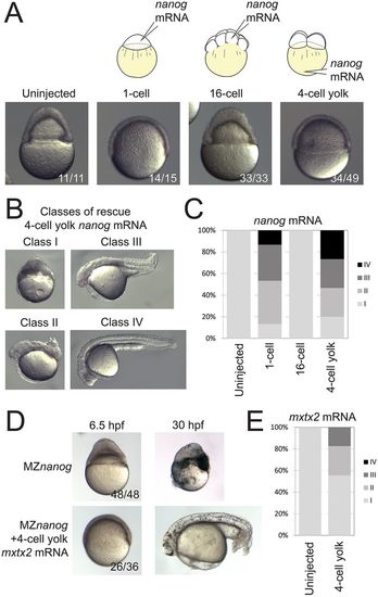
Rescue of MZnanog embryos by YSL expression of nanog or mxtx2 mRNA. (A) Uninjected MZnanog embryos, or MZnanog embryos injected with 33 pg GFP mRNA and 25 pg nanog mRNA into the cell at the 1-cell stage, into a cell at the 16-cell stage, or into the vegetal yolk at the 4-cell stage, were sorted for appropriate spatial GFP expression at sphere stage (as shown in Fig. S1A), and scored and imaged at 8 hpf. (B) Embryos were scored at 24 hpf into four rescue phenotype classes as described in Materials and Methods. (C) Quantification of rescue phenotype scores illustrated as stacked bar plots (uninjected n=11; 1-cell n=15; 16-cell n=33; 4-cell yolk n=30). (D,E) Control MZnanog embryos, or embryos injected into the vegetal yolk at the 4-cell stage with 33 pg GFP mRNA and 50 pg mxtx2 mRNA, were sorted for appropriate spatial GFP expression at sphere stage, scored as above, and imaged at 6.5 hpf and at 30 hpf (D). These scores were quantified and illustrated as stacked bar plots (E) (control n=48; 4-cell yolk n=92). In A and D, the number of embryos exhibiting the illustrated phenotype is shown in the bottom-right corner of each image as a proportion of total embryos examined. |