
Fig. 5
- ID
- ZDB-FIG-180426-4
- Publication
- Ji et al., 2017 - Involvement of Lypge in the formation of eye and pineal gland in zebrafish
- Other Figures
- All Figure Page
- Back to All Figure Page
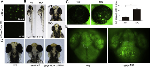
Knockdown of lypge causes small eye and small head phenotype. (A) Examination of lypge MO efficiency, left: expression of EGFP fluorescence in embryos injected MO-binding site + EGFP mRNA alone. Right: expression of EGFP fluorescence is absent from embryos co-injected MO-binding site + EGFP mRNA with lypge MO. (B) Compared with phenotype between wildtype (WT) embryos and morphants (MO) at 52 hpf. (C) Cell apoptosis assay, embryos are stained with acridine orange (AO) at 48 hpf and quantification of the number of AO positive cells in eye (green). Statistically significant differences between the WT and MO (Fisher's t-test) are indicated: ***P < 0.001. Error bars indicate s.e.m. (D) compared with phenotype in wildtype, lypge MO injected and lypge MO and p53 MO co-injected embryos at 76 hpf. (E) Apoptosis of brain cells in WT embryos and morphants. Region of pineal gland is indicated by white dotted line. (For interpretation of the references to colour in this figure legend, the reader is referred to the web version of this article.) |
| Fish: | |
|---|---|
| Knockdown Reagents: | |
| Observed In: | |
| Stage: | Protruding-mouth |
Reprinted from Gene, 642, Ji, D., Wang, S., Li, M., Zhang, S., Li, H., Involvement of Lypge in the formation of eye and pineal gland in zebrafish, 491-497, Copyright (2017) with permission from Elsevier. Full text @ Gene