
Fig. 5
- ID
- ZDB-FIG-180424-17
- Publication
- Silvent et al., 2017 - Zebrafish skeleton development: High resolution micro-CT and FIB-SEM block surface serial imaging for phenotype identification
- Other Figures
- All Figure Page
- Back to All Figure Page
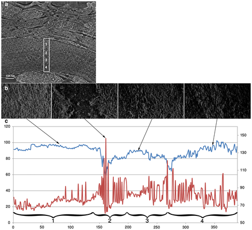
Directionality and dispersion analyses of the collagen fibrils in the tail bone at 30 dpf. a. FIB slice of the demineralized bone, observed in transverse plane. The rectangle spans the bone thickness. b. FIB slices characteristic of the different regions observed in the rectangle, observed in frontal plane, Slice 80 for region 1, slice 161 for region 2, slice 216 for region 3 and slice 346 for region 4. c. Direction (in blue) and dispersion (in red) plots of the collagen fibrils for each slice in the stack. The left vertical axis shows the azimuthal direction angle whereas the right shows the angular dispersion. |