
Fig. 3
- ID
- ZDB-FIG-180418-25
- Publication
- Ando et al., 2017 - Osteoblast Production by Reserved Progenitor Cells in Zebrafish Bone Regeneration and Maintenance
- Other Figures
- All Figure Page
- Back to All Figure Page
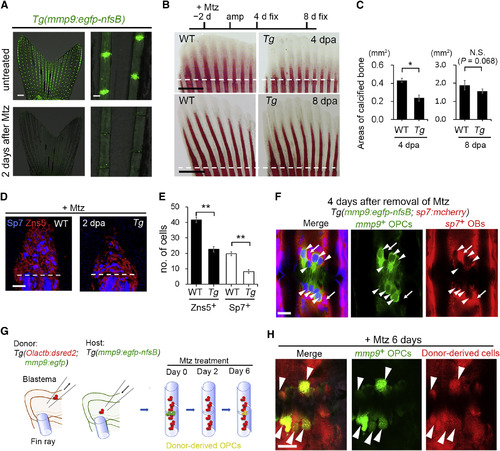
OPCs Replenished from Mesenchymal Precursors Are Significant Contributors to Bone Regeneration (A) A representative example of OPC ablation. Effective ablation of the OPCs occurs with 5 mM Mtz treatment for 2 days. Scale bars, 500 μm (left panel) and 50 μm (right panel). (B) Alizarin red S staining of regenerating WT and nfsB-expressing Tg fins treated with Mtz. Dashed lines indicate fin amputation sites. Scale bars, 500 μm. (C) Quantification of calcified areas in (B). A significant decrease of calcified tissue was observed when OPCs were ablated. Error bars indicate mean ± SEM. ∗p < 0.01, Student's two-tailed t test; n = 5 fins. N.S., not significant. (D) Detection of Zns5 and Sp7 in WT and OPC-ablated Tg regenerates. Dashed lines indicate fin amputation sites. Nuclei were counterstained with DAPI. Scale bars, 10 μm. (E) Quantification of (D). The numbers of Zns5+ and Sp7+ cells, respectively, were significantly decreased by OPC ablation. Error bars indicate mean ± SEM. ∗∗p < 0.001, Student's two-tailed t test; n = 6 confocal optical sections from different fin rays (total 5 fish). (F) Replenishment of OPCs from non-osteoblast precursors. In double Tg(sp7:mcherry; mmp9:egfp-nfsB), re-formed OPCs (arrowheads) after ablation were not mCherry+ (n = 15 of 15 joints from total 5 fish), indicating that re-formed OPCs were not derived from mCherry+ osteoblasts. Arrows point to nearby osteoblasts. Nuclei, DAPI. Scale bar, 10 μm. (G) Procedure of mesenchymal cell transplantation and host OPC ablation. Olactb:dsRed2 refers to Olactb:loxP-dsred2-loxP-egfp. Donor blastema was transplanted into the host blastema region (Shibata et al., 2016). Most of the transplanted cells contribute to mesenchymal cells. Eight days after transplantation, host OPCs were ablated with 5 mM Mtz to see whether or not the re-formed joint cells (EGFP+) were derived from DsRed2+ mesenchymal cells. (H) Emergence of OPCs from mesenchymal cells. Arrowheads point to re-formed OPCs derived from DsRed2+ mesenchyme. n = 24 fin ray joints from total 5 fish. Scale bar, 10 μm. |
Reprinted from Developmental Cell, 43(5), Ando, K., Shibata, E., Hans, S., Brand, M., Kawakami, A., Osteoblast Production by Reserved Progenitor Cells in Zebrafish Bone Regeneration and Maintenance, 643-650.e3, Copyright (2017) with permission from Elsevier. Full text @ Dev. Cell