
FIGURE
Fig. S1
- ID
- ZDB-FIG-180404-47
- Publication
- Liu et al., 2017 - Overexpression of DYRK1A, a Down Syndrome Candidate gene, Impairs Primordial Germ Cells Maintenance and Migration in zebrafish
- Other Figures
- All Figure Page
- Back to All Figure Page
Fig. S1
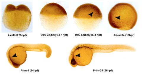
Spatiotemporal expression pattern of Dyrk1a protein in zebrafish embryos. Detection of zebrafish Dyrk1a protein using whole-mount immunohistochemistry (WIHC) at indicated stages. Arrows show the stronger expression region of Dyrk1a protein. It is shown that expression pattern of Dyrk1a protein is similar to dyrk1a transcripts detected by WISH. Embryo orientations: 2-cell, 30%-epiboly and 50%-epiboly stage, lateral views with the animal pole oriented at the top; 8-somite, Prim-5 and Prim-25 stage, lateral views with anterior oriented toward the left. |
Expression Data
| Antibody: | |
|---|---|
| Fish: | |
| Anatomical Terms: | |
| Stage Range: | 2-cell to Prim-25 |
Expression Detail
Antibody Labeling
Phenotype Data
Phenotype Detail
Acknowledgments
This image is the copyrighted work of the attributed author or publisher, and
ZFIN has permission only to display this image to its users.
Additional permissions should be obtained from the applicable author or publisher of the image.
Full text @ Sci. Rep.