
Fig. 5
- ID
- ZDB-FIG-180404-35
- Publication
- Gonzalez-Nunez, 2015 - Role of gabra2, GABAA receptor alpha-2 subunit, in CNS development.
- Other Figures
- All Figure Page
- Back to All Figure Page
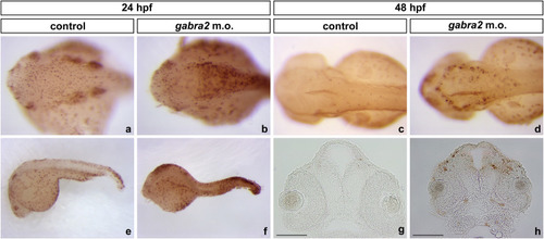
Apoptosis assays on gabra2 morphant embryos. TUNEL staining on gabra2 deficient embryos of 24 hpf (a and b – dorsal views; e and f – lateral views) and 48 hpf (c and d – dorsal views; g and h – 20 μm thick coronal sections). At 24 hpf, there is an increase in the number of apoptotic nuclei (which are positive for peroxidase labelling) in the hindbrain region of morpholino-injected embryos [69.30±15.52 apoptotic nuclei in control embryos as compared to 164.6±36.87 in morphant embryos; * p<0.05 as determined by one sample t-test]. Apoptosis is reduced at 48 hpf when compared to morphants at 24 hpf (* p<0.05 as determined by one sample t-test), although gabra2 deficient fish display apoptotic cells in the walls of the third ventricle and in some regions of the forebrain. [36.87±4.67 apoptotic nuclei in control embryos as compared to 77.06±5.34 in morphant embryos; ** p<0.01 as determined by one sample t-test]. (a–f) Embryos are oriented anterior towards the left and posterior towards the right. (g and h) Orientation of embryos is anterior up; scale bar: 100 μm. |
| Fish: | |
|---|---|
| Knockdown Reagent: | |
| Observed In: | |
| Stage Range: | Prim-5 to Long-pec |