
Fig. s1
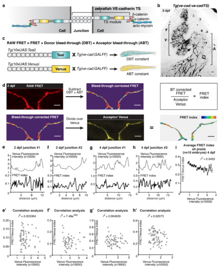
Controlled FRET quantification of VE-cadherin-TS expression (a) Schematic representation of VE-cadherin Tension Sensor (TS) proteins at a cell-cell junction and intra-cellular linkage to acto-myosin (blue) via β-catenin (red) and α-catenin (green). (b) Venus expression (grey) from Tg(ve-cad:ve-cadTS) in the endocardium of the heart at 3 dpf (A=atrial endocardium, V=ventricular endocardium). Scale bar = 10μm. (c) Schematic representation of Tg(10xUAS:Teal) and Tg(10xUAS:Venus) strains required to correct for Donor bleed through (DBT) and Acceptor bleed through (ABT). (d) Step-wise explanation of RAW FRET processing. From the intensity of each pixel in the RAW FRET image the value for DBT and ABT is subtracted, resulting in the bleed through (BT) corrected FRET. The BT-corrected FRET is subsequently divided by the intensity for Venus in each pixel to normalise for the amount of protein present. The final outcome is represented as a ratio-metric FRET. Scale bar = 5μm. (e-f’) Intensity plots of Venus fluorescence (grey, top) and FRET index (black, below) from spatially matched pixels within a line drawn across single junctions at 2 dpf (e-f) showing that these quantities to not follow the same profile. Correlation analysis of matched pixels within these junctions is depicted in e’ (r2=0.005305, p=0.5491) and f’ (r2=7.458e-005, p=0.9487). (g-h’) Intensity plots of Venus fluorescence (grey, top) and FRET index (black, below) from spatially matched pixels within a line drawn across single junctions at 4 dpf (g-h) showing that these quantities to not follow the same profile. Correlation analysis of matched pixels within these junctions is depicted in g’ (r2=0.004929, p=0.5694) and h’ (r2=0.02073, p=0.1941). (i) Grouped analysis of ratio-metric FRET values in all pixels within n=52 junctional ROIs selected from n=10 embryos at 4 dpf. Pixels were grouped based on increments of Venus intensity levels of 500. The mean FRET-index value and S.E.M of each Venus intensity group was calculated and plotted. Correlation analysis of mean Venus intensity and mean FRET index within each group (r2=0.5453, ****p<0.0001). |