
Fig. 2
- ID
- ZDB-FIG-180403-11
- Publication
- Posner et al., 2017 - The zebrafish as a model system for analyzing mammalian and native α-crystallin promoter function.
- Other Figures
- All Figure Page
- Back to All Figure Page
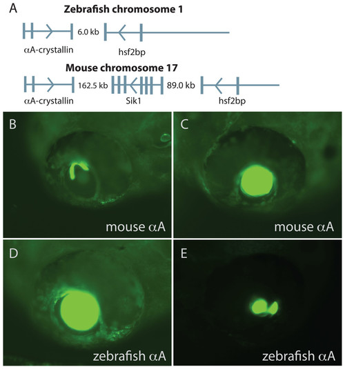
Comparison of mouse and zebrafish αA-crystallin chromosomal arrangement and their ability to drive GFP expression in zebrafish embryos. The structural and functional conservation of mammalian and zebrafish αA-crystallin is mirrored in their shared syntenic relationship with hsf2bp (A). Vertical bars note exons, thin horizontal lines note introns and arrows show direction of transcription. The promoter regions for each gene produced similar temporal and spatial expression patterns (B–E), with expression almost exclusively restricted to the lens. The extent of lens expression varied for both orthologous promoters (compare B to C for mouse and D to E for zebrafish). |