
FIGURE
Fig. 1
- ID
- ZDB-FIG-180403-10
- Publication
- Posner et al., 2017 - The zebrafish as a model system for analyzing mammalian and native α-crystallin promoter function.
- Other Figures
- All Figure Page
- Back to All Figure Page
Fig. 1
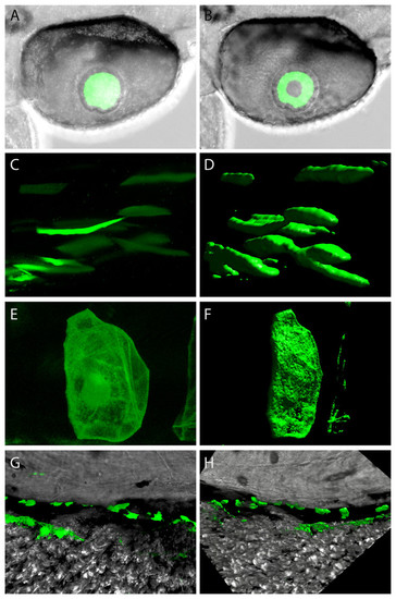
Confocal imagery showing representative sites of GFP expression produced by mouse and zebrafish α-crystallin promoters. Examples of lens expression produced with a zebrafish αA promoter (A and B). Various sites of extraocular expression shown as single z-planes (on left) and as 3-dimensional renders (on right) for skeletal muscle produced with a mouse αB promoter (C and D); for notochord produced with a zebrafish αBb promoter (E and F); dorsal to the yolk produced with a zebrafish aA promoter (G and H). |
Expression Data
Expression Detail
Antibody Labeling
Phenotype Data
Phenotype Detail
Acknowledgments
This image is the copyrighted work of the attributed author or publisher, and
ZFIN has permission only to display this image to its users.
Additional permissions should be obtained from the applicable author or publisher of the image.
Full text @ Peer J.