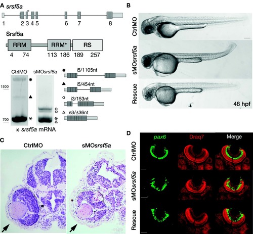
Injection of a splice site blocking MO targeting the srsf5a gene led to developmental defects. (A) srsf5a is composed of eight exons. The protein is encoded by exons 2–8 and consists of two RRM domains responsible for RNA binding and one RS domain, essential for protein–protein interactions. Three different transcripts are produced from srsf5a. Among them, two alternative transcripts retain intron 5 or a part of it (black up-pointing triangle and black dot). sMOsrsf5a was designed to target the exon3–intron3 junction. RT-PCR experiments to amplify srsf5a mRNA in control and morphant embryos at 48 hpf revealed intron3 retention (open hexagon mark), introducing a premature STOP codon into the RRM1 encoding part of the mRNA. Expression of the three normal srsf5a transcripts was strongly reduced in morphants, while MO injection also triggered the use by the splicing machinery of a cryptic splice site located in exon3, and leading to a deletion of 36 bases in the open reading frame of the srsf5a transcript (srsf5a/Δ36, open triangle mark). The resulting protein has a 12-amino acids deletion within the RRM1. The open square mark corresponds to a srsf5a transcript in which intron3 is retained and with a deletion of 36 bases in the exon3 (srsf5a/Δ36-intron3). All PCR products were identified by sequencing. (B) Zebrafish embryos injected with 3 ng of ctrlMO or sMOsrsf5a with or without a rescuing dose of srsf5a mRNA (80 pg) at 48 hpf. The defects in brain, eye and curved tail could be partially rescued by srsf5a mRNA injection. Pigmentation was not visible as embryos were treated with 1-phenyl-2-thiourea to increase their transparency. Bar: 200 μm. (C) Haematoxylin/eosin sections obtained from ctrlMO and sMOsrsf5a injected embryos revealed abnormal organization of cells in the retina and an increase of cell death in the eye and the entire brain at 48 and 72 hpf (data not shown). (D) Fluorescent in situ hybridization using a pax6b probe followed by nuclear staining using draq7® revealed the disorganization of the ganglional cell layer and of the inner nuclear layer in the retina at 72 hpf in morphants compared to control embryos. Rescue experiments allowed us to partially restore the control phenotype. Scale bar: 50 μm.
|
