
Fig. 7
- ID
- ZDB-FIG-180103-29
- Publication
- Lu et al., 2017 - Ablation of EYS in zebrafish causes mislocalisation of outer segment proteins, F-actin disruption and cone-rod dystrophy
- Other Figures
- All Figure Page
- Back to All Figure Page
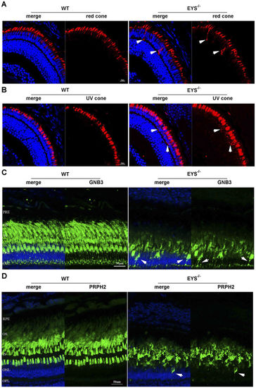
Photoreceptor OS proteins were mislocalised in EYS−/− zebrafish. (A) Retinal cryosections from WT and EYS−/− zebrafish were immunostained with anti-opn1LW antibodies at 10 dpf. White arrows indicated the mislocalised opn1LW proteins. Scale bars: 10 μm. (B) Retinal cryosections from WT and EYS−/− zebrafish were immunostained with anti-opn1SW1 antibodies at 10 dpf. White arrows indicated the mislocalised opn1SW1 proteins. Scale bars: 10 μm. (C) Retinal cryosections from WT and EYS−/− zebrafish were immunostained with anti-GNB3 antibodies at the age of 5 mpf. White arrows indicate the mislocalised GNB3 proteins. Scale bars: 50 μm. (D) Retinal cryosections from WT and EYS−/− zebrafish were immunostained with anti-PRPH2 antibodies at the age of 7 mpf. White arrows indicated the mislocalised PRPH2 proteins. Scale bars: 50 μm. |
| Antibodies: | |
|---|---|
| Fish: | |
| Anatomical Terms: | |
| Stage Range: | Days 7-13 to Adult |
| Fish: | |
|---|---|
| Observed In: | |
| Stage Range: | Days 7-13 to Adult |