
Fig. 5
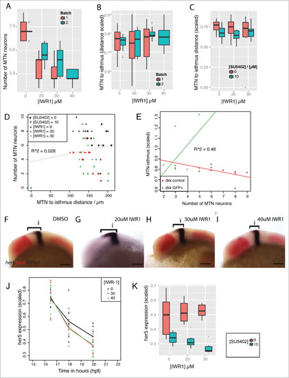
Tankyrase activity does not affect positioning of MTN neurons or FGF directed her5 expression in the dorsal midbrain. Box plot of the number of MTN neurons (A) or MTN-isthmus distance (scaled by midbrain size, B) in 2 separate clutches of embryos (red, blue) treated with 0, 20, 30, 40mM IWR-1 from 14–24 hpf (n = 10 for each condition). Box plot of MTN-isthmus distance (scaled) in 24 hpf embryos treated with DMSO (red) or 10mM SU5402 (blue) in conjunction with IWR-1 (C, n = 10 for each condition). Dot plot of MTN neuron number relative to MTN-isthmus distance (scaled) for embryos treated with IWR-1 (0, 20, 30, 40mM) and SU5402 (0, 10mM) from 14 hpf (D). A correlation test reveals no correlation between neuron number and distance (R = 0.028, n = 10 for each condition). Dot plot of MTN-isthmus distance (scaled) relative to MTN number in 24 hpf control embryos (red) or Tg[hsp70l:dkk1b-gfp] embryos expressing dkk1b (green) from 16.5 hpf (E). A correlation test reveals no correlation between MTN neuron number and distance (R = 0.48, n = 10 for each condition). Lateral views of 18 hpf embryos processed by in situ hybridization to reveal expression of her5 (blue, bracket indicates expression extent in dorsal midbrain) and pax6 (red) following treatment with IWR-1 at 0, 20, 30, 40mM IWR-1 from 14 hpf (F-I). A line plot of the her5 expression domain in the dorsal midbrain (scaled to midbrain size) in embryos treated with IWR-1 at 0, 30, 40mM from 14 hpf at 16.5, 18, 20 hpf (J, n = 10 for each condition). Box plot of her5 expression domain in the dorsal midbrain (scaled) in 24 hpf embryos treated with IWR-1 at 0, 20, 30mM and SU5402 at 0 (red) and 10mM (blue) from 14 hpf (K, n = 10 for each condition). Isthmus (i). Scale bars: 100mm. |