
Fig. 1
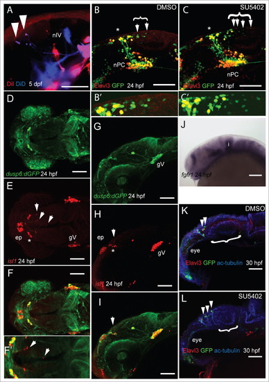
FGF signaling dictates the spatial positioning of MTN neurons in the midbrain during development. DiI (red) labeling of masseter and and DiD (blue) labeling of levator arcus palatini and lateral rectus cranial muscles in 5 dpf larvae leads to retrograde labeling of MTN neurons in the anterior midbrain (A). Lateral views of Tg[elavl3:egfp] embryos labeled with anti-GFP (green) and anti-Elavl3 (HuC/D, red) reveals the presence of more MTN neurons (arrowheads) at posterior locations in the midbrain following treatment with 40mM SU5402 from 14–24 hpf (C) relative to DMSO exposure (B). Bracket indicates extent of MTN neurons (represented by enlarged area in B' and C'). Dorsal (D–F) and lateral (G–I,) views of 24 hpf Tg [dusp6:d2eGFP] embryos processed with anti-GFP (green) and anti-Isl1 (red). A zoomed dorsal view (F) reveals that neurons of the nucleus of the tract of the posterior commissure (nTPC, asterisk) and MTN neurons (arrowheads) develop in regions devoid of anti-GFP labeling. At 24 hpf, fgfr1 expression is absent from the anterior midbrain (J). Lateral views of 30 hpf Tg[dlx5a/6a:eGFP] embryos labeled with anti-Elavl3 (red), anti-acetylated tubulin (blue) and anti-GFP (green) reveals more MTN neurons (arrowheads) and a reduced optic tectum (bracket) following exposure to 40mM SU5402 (L) from 14–24 hpf compared to DMSO treated control animals (K). Nucleus of the posterior commissure (nPC), trochlear nerve (nIV). Scale bars: 50mm (A), 100mm (B–L). |