
FIGURE
Fig. S2
- ID
- ZDB-FIG-171201-26
- Publication
- Vliegenthart et al., 2017 - Characterization of Triptolide-Induced Hepatotoxicity by Imaging and Transcriptomics in a Novel Zebrafish Model
- Other Figures
- All Figure Page
- Back to All Figure Page
Fig. S2
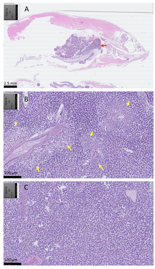
Histological images of adult zebrafish after exposure to triptolide (1.6 μM) for 10 hours. (A) Subgross view. Liver is indicated by the red arrow. The area displaying hepatic necrosis is indicated by the circle. (B) Arrows denote lesions consistent with hepatic necrosis after triptolide exposure. (C) Control adult zebrafish after exposure to vehicle control. Normal liver, with no areas of necrosis. |
Expression Data
Expression Detail
Antibody Labeling
Phenotype Data
Phenotype Detail
Acknowledgments
This image is the copyrighted work of the attributed author or publisher, and
ZFIN has permission only to display this image to its users.
Additional permissions should be obtained from the applicable author or publisher of the image.
Full text @ Toxicol. Sci.