
Fig. S8
- ID
- ZDB-FIG-171113-3
- Publication
- Sánchez et al., 2016 - Mechanosensory organ regeneration in zebrafish depends on a population of multipotent progenitor cells kept latent by Schwann cells
- Other Figures
- All Figure Page
- Back to All Figure Page
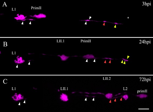
INCs respond to damage and behave like multipotent progenitor cells. Blastula cells from Tg(ubiquitin:RFP) embryos transplanted into tg(et20:GFP) embryos result in chimeric larvae with a few labeled INCs (magenta, labeled with arrowheads) that can be followed through time in vivo. (A) At 3 dpf, a chimeric larva was electroablated in the L2 neuromast; the asterisk shows the injury site. A proximal (yellow arrowhead) and more distal (red and white arrowheads) labeled INCs can be observed. (B) After 24 hpi, the INC proximal to the injury zone has proliferated and daughter cells begin to accumulate (yellow arrowheads) whereas the nearest neighboring INC seen in A divides once but the daughter cells do not move from their position (red arrowheads). The farthest labeled INCs (white arrowheads) do not divide. (C) After 72 hpi, a regenerated L2 neuromast appears in the position where the injury was made. With the exception of two cells, the entire regenerated L2 neuromast is derived from the single labeled INC cell (yellow arrowhead in A). The distal INC has divided once more but does not contribute to the regenerated neuromast (red arrowheads). From 24 hpi to 72 hpi (which corresponds to 4–6 dpf), the PrimII travels through this region and deposits the secondary neuromasts LII.1 and LII.2. Scale bar: 50 μm. (TIF 1069 kb) |