
Fig. S2
- ID
- ZDB-FIG-171110-43
- Publication
- Sánchez et al., 2016 - Mechanosensory organ regeneration in zebrafish depends on a population of multipotent progenitor cells kept latent by Schwann cells
- Other Figures
- All Figure Page
- Back to All Figure Page
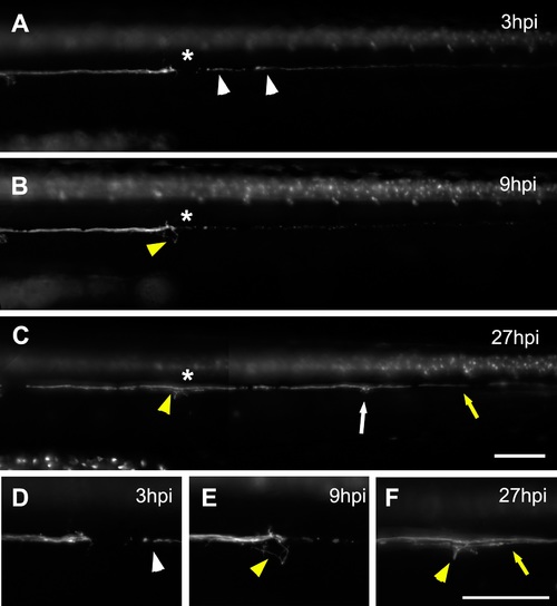
Behavior of the lateral line nerve after neuromast electroablation. g(neurod:GFP; cxcr4b:mCherry) larvae that express mCherry in all cells types of the lateral line and GFP in the lateral line nerve were electroablated at 3 dpf in the L3 neuromast (asterisk); images show only the GFP channel. (A) At 3 hpi, degenerating fragments of denervated axons are still present at the myoseptum (white arrowheads). (B) At 9 hpi, nerve fragments have been cleared and axons sprouting from the nerve stump begin to extend and explore the injured region (yellow arrowhead). (C) From 9 to 27 hpi, the nerve regrows caudally (yellow arrow). The asterisks indicates the injury site and the yellow arrowhead shows axons innervating this region. Also, the regrowing axons innervate more caudal neuromasts (white arrow). (D–F) Higher magnification of the injury zone at 3 hpi (D), 9 hpi (E), and 27 hpi (F). Scale bar: 100 μm. |