
Fig. 3
- ID
- ZDB-FIG-171006-7
- Publication
- Xiyuan et al., 2017 - NO-sGC Pathway Modulates Ca2+ Release and Muscle Contraction in Zebrafish Skeletal Muscle.
- Other Figures
- All Figure Page
- Back to All Figure Page
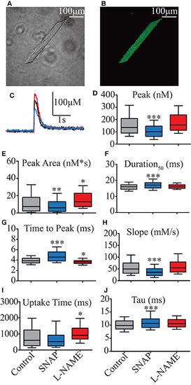
Effect of NO on biophysical parameters of isolated zebrafish myocyte Ca2+ transients. (A) Photomicrography of isolated zebrafish myocytes under light microscope. (B) Photomicrography of the same myocyte shown in (A) loaded with 10 μM Fluo4-AM and seen under fluorescence microscope. (C) Representative Ca2+ transient's traces obtained from electrical field stimulation. Control represented in black, 00 μM SNAP in blue, and 5 mM L-NAME in red. Statistical analyses of Ca2+ transients' biophysical parameters: (D) Peak (nM), (E) Duration50 (ms), (F) Peak Area (nM*s), (G) Time to Peak (ms), (H) Slope (μM/s), (I) Uptake Time (ms), and (J) Tau (s). Values are expressed as median; 25–75%. *p < 0.05, **p < 0.01, ***p < 0.001 vs. control; n = 278 cells for control, 153 cells for SNAP and 106 cells for L-NAME. |