
- Title
-
NO-sGC Pathway Modulates Ca2+ Release and Muscle Contraction in Zebrafish Skeletal Muscle.
- Authors
- Xiyuan, Z., Fink, R.H.A., Mosqueira, M.
- Source
- Full text @ Front. Physiol.
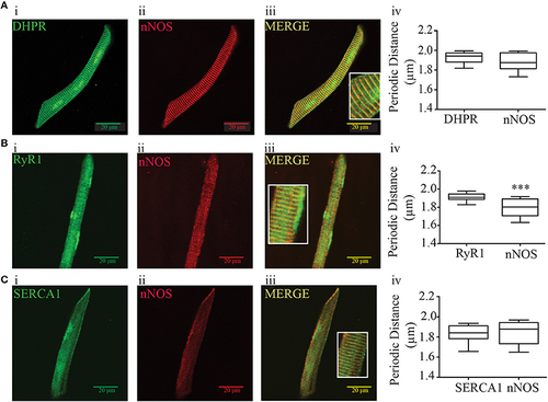
Immunofluorescence and periodic distance analysis of DHPR, RyR, SERCA, and nNOS from zebrafish isolated myocytes. (A) Immunofluorescence for DHPR (i), nNOS (ii), and merge (iii), and periodic distance analysis (iv) from 26 cells. (B) Immunofluorescence for RyR (i), nNOS (ii), and merge (iii), and periodic distance analysis (iv) from 18 cells. (C) Immunofluorescence for DHPR (i), nNOS (ii), and merge (iii), and periodic distance analysis (iv) from 16 cells. The insert at the merged picture shows a magnification of the respective picture. Values are expressed as median; 25–75%. ***p < 0.001 vs. RyR immunofluorescence. |
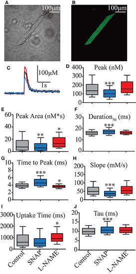
Effect of NO on biophysical parameters of isolated zebrafish myocyte Ca2+ transients. (A) Photomicrography of isolated zebrafish myocytes under light microscope. (B) Photomicrography of the same myocyte shown in (A) loaded with 10 μM Fluo4-AM and seen under fluorescence microscope. (C) Representative Ca2+ transient's traces obtained from electrical field stimulation. Control represented in black, 00 μM SNAP in blue, and 5 mM L-NAME in red. Statistical analyses of Ca2+ transients' biophysical parameters: (D) Peak (nM), (E) Duration50 (ms), (F) Peak Area (nM*s), (G) Time to Peak (ms), (H) Slope (μM/s), (I) Uptake Time (ms), and (J) Tau (s). Values are expressed as median; 25–75%. *p < 0.05, **p < 0.01, ***p < 0.001 vs. control; n = 278 cells for control, 153 cells for SNAP and 106 cells for L-NAME. |
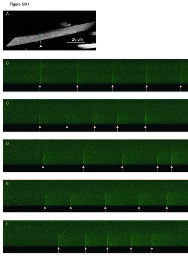
Ca2+ transients do not occur in the entire isolated myocyte of 5-7 dpf zebrafish. A. Photomicrography of isolated myocyte of zebrafish larvae indicating with an arrowhead the position of the line-scanning. B-F. Line-scanning from the sub-sarcolemmal level of one side of the myocyte until the other with one micrometer step. In each level, several electrical stimulation was applied as indicated by the white arrows. |