
FIGURE
Fig. S1
- ID
- ZDB-FIG-170908-16
- Publication
- Breau et al., 2017 - Extrinsic mechanical forces mediate retrograde axon extension in a developing neuronal circuit
- Other Figures
- All Figure Page
- Back to All Figure Page
Fig. S1
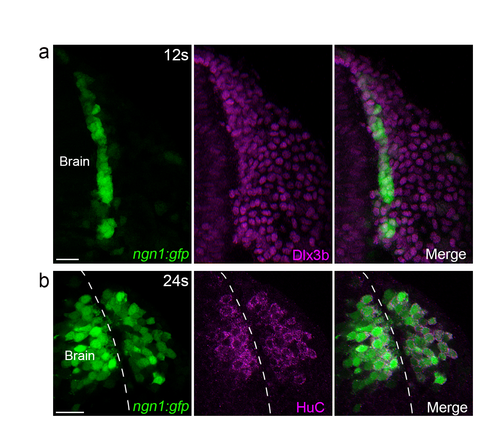
Characterisation of the ngn1:gfp transgenic line in the head during OP coalescence. (a) Dlx3b immunostaining performed on a ngn1 :gfp embryo at 11-12s. GFP (green) is expressed in a narrow cell domain flanking the anterior brain and expressing Dlx3b (magenta), a marker for OP cells. (b) Hue immunostaining (magenta) performed on a ngn1 :gfp embryo at 24s, showing co-localisation between GFP and Hue. Scale bars: 25 µm. |
Expression Data
Expression Detail
Antibody Labeling
Phenotype Data
Phenotype Detail
Acknowledgments
This image is the copyrighted work of the attributed author or publisher, and
ZFIN has permission only to display this image to its users.
Additional permissions should be obtained from the applicable author or publisher of the image.
Full text @ Nat. Commun.